It is the most widely used and the most important staining technique in bacteriology especially in medical bacteriology. There are two types of bacteria based on their cell wall composition- Gram-positive and gram-negative.

Lab Midterm Lab 3 Flashcards Quizlet
Both gram-positive and gram-negative cells have.
. Escucha la conversación entre daniel y. If the bacteria turns pink or red they are Gram-negative. After the addition of the counterstain in gram staining the gram-positive bacterial cell appears purple whereas the gram-negative one appears pink.
The bacteria are differentiated through a series of staining and decolorization steps. If the bacteria stays purple they are Gram-positive. In Grams method which is based on the ability of a cell in retaining the crystal violet dye during solvent treatment it is the difference in the microbial cell wall that is amplified.
To make the flagella visible. If you dont proactively choose a different repayment plan option your federal student loans will default to the standard plan which has a term of. To make the bacterial cells larger.
A Gram stain is colored purple. The slide is stained with safranin for 1min then rinse with water very gently until no color is coming from the water. Textbook Solutions Expert Tutors Earn.
Practical 1 study questions Cells are differentiated after which step in the Gram. Cells are differentiated after which step in gram stain. The cell walls for gram-negative microorganisms have a higher lipid content than gram-positive cells.
Water tap water or deionized 6. Since human cells do not have cell walls or peptidoglycan the gram stain would do nothing because the primary stain would wash out notes Wikipedia. The organisms that do not take up primary stain appear red under a microscope and are Gram-negative organisms.
The purpose of a mordant in the Gram stain is To remove the simple stain. First cells are stained with crystal violet followed by the addition of a. The Gram stain involves staining bacteria fixing the color with a mordant decolorizing the cells and applying a counterstain.
B The best use of a negative stain is. Supplies and Reagents 1. The four basic steps of the Gram Stain are.
The gram-positive bacteria will stain dark blue or purple. The procedure is based on the reaction between peptidoglycan in the cell walls of some bacteria. The process of gram staining is used to differentiate between gram-positive and gram-negative bacteria notes Sciences 360.
Acetone alcohol is the decolorizer. A To see endospores. The gram-negative bacteria will stain pink.
Place the following steps in the correct sequence. Place the slide on the microscope to view the sample. It removes the primary stain crystal violet from the cell wall of Gram negative cells allowing them to.
The primary stain crystal violet binds to peptidoglycan coloring cells purple. When the stain combines with bacteria in a sample the bacteria will either stay purple or turn pink or red. CV dissociates in aqueous solutions into CV and Cl ions.
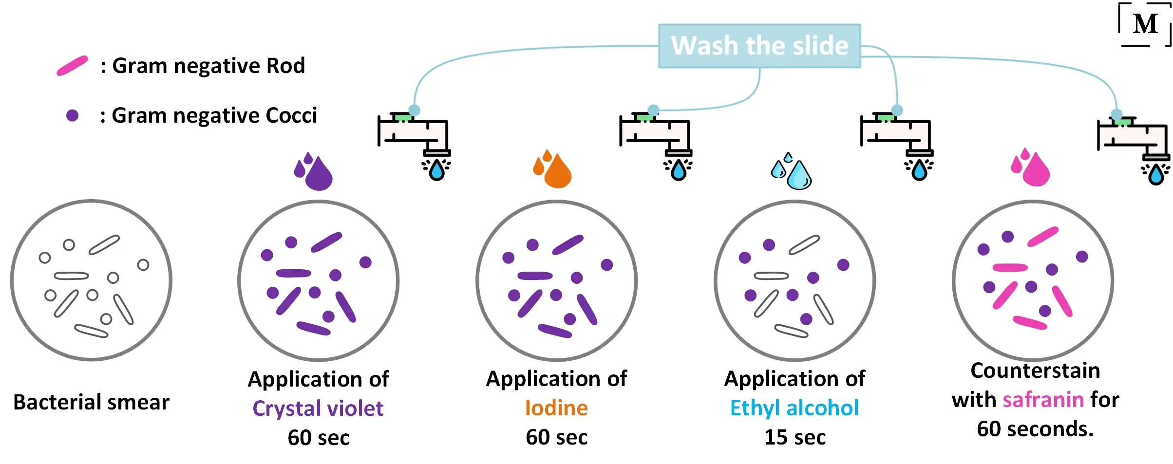
1 Application of the primary stain Crystal Violet CV to a heat-fixed smear of bacterial culture. After step one all cells will look violet Step 2 of gram slide flooded with iodine mordant for 1 min then rinsed with water After step 2 all cells will look violet step 3 of gram slide rinsed with solution of ethanol and acetone decolorizer for 10-30 sec then rinsed with water after step 3. To make gram-negative cells visible.
Gram staining is a differential bacterial staining technique used to differentiate bacteria into Gram Positive and Gram Negative types according to their cell wall composition. Explain the difference between the constitutional and informal requirements to become president. To prevent the crystal violet from leaving the cells.
E The order doesnt matter. The first step in gram staining is the use of crystal violet dye for the slides initial staining. Gram staining involves four steps.
As a differential staining technique the gram staining procedure distinguishes bacterial cells based on the properties of their cell wall. These differences affect many aspects of the cell including the way the cell takes up and retains stains. That said I have been know to make a supposition based on experience of various organisms being seen often under the mic.
Gram staining targets the cell wall and a layer called peptidoglycan. Gram positive cells take up the crystal violet which is then fixed in the cell with the iodine mordant. The expulsion of the lipid layer improves the draining of the primary stain from the cells into the surrounding solvent.
The two categories cause different types of infections. The difference is attributed to the differences in their cell wall. Blot dry with absorbent paper.
Gram-positive infections include methicillin-resistant Staphylococcus aureus strep. Personal protective equipment 2. Upload your study docs or become a Course Hero member to access this document Continue to access End of preview.
Because of the way the gram stain works I would say there must be all elements of the stain present to be able to absolutely identify the gram result of an organism. The differential nature of the Gram stain is based on the ability of some bacterial cells to retain a primary stain crystal violet by resisting a decolorization process. Absorbent paper such as bibulous paper 5.
The next step also known as fixing the dye involves using iodine to form crystal violet- iodine complex to prevent easy removal of dye. Gram negative and gram positive organisms are distinguished from each other by differences in their cell walls. Gram-positive cells will stain purple and Gram-negative cells will stain red to pink.
These two ions then penetrate through the cell wall and cell membrane of both Gram-positive and Gram-negative cells.
Gram Staining Better Understanding Of The Procedure And Easy Interpretation Of The Results

Pin Em Medimoon Com

Development Of A Standardized Gram Stain Procedure For Bacteria And Inflammatory Cells Using An Automated Staining Instrument Li 2020 Microbiologyopen Wiley Online Library


0 Comments